


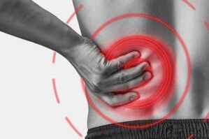
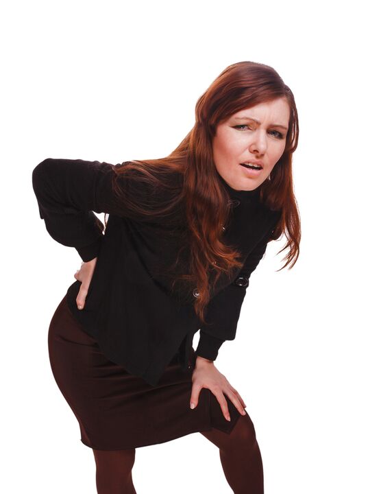
Lumbar osteochondria is a chronic degenerative dystrophic disease of the lumbar spine that affects the structure of the intervertebral discs and a number of lumbar spine.It affects her people primarily work.It manifests in various symptoms, the main ones of which are lower back pain and legs, limiting movements on the lower back.For diagnostics, research methods such as x -ray, computed tomography or magnetic coordination of the lumbar spine are used.In this article, you can more detailed with the causes, symptoms and methods of diagnosis of lumbar spine.
Osteochondria is the result of body aging.These or other signs of this disease can be found in almost every person (!), Starting at 25 years.But here is the seriousness of these changes, the pace of their evolution, the degree of clinical events depends on many causes, mainly on how a healthy lifestyle leads a particular person.Moderate physical exercise, compulsory morning exercise, proper posture of the body when a series of work is performed (garden, construction, dangerous cleaning of the house and so on)
According to statistics, spine osteochondria in 80% of cases is the cause of back pain.

How does osteochondria grow?
The entire spine consists of separate vertebrae, between the bodies of which there are intervertebral discs.That is, between the two vertebrae is a disc.The disc consists of gelatinous (polyp) core and fibrous ring.The core contains a lot of water and provides spine depreciation and flexibility.The fibrous ring is located along the periphery of the core of the jacket, as if it was holding it inside.
With a prolonged increased load at the pier core, it changes its normal properties, loses water and dries the sequences: the disc is flattened and the vertebral bodies approach each other.Along with such procedures, in the core jacket, the fibrous ring loses its elasticity and, under the influence of mechanical loads, begins to protrude.This is called a protrusion.Then the fibrous ring cracks and a gelatinous core falls through the resulting gaps: a disc hernia appears.A graph of two adjacent vertebrae and a disc located between them, called the vertebral section, acquires excessive mobility, thereby increasing the load on the nearby sections.Overloading adjacent parts causes a similar pathological process in them.These changes are called osteochondria.
In order to somehow ensure the stability of the spine, bone increases are formed along the edges of the vertebral bodies, increasing the support area.This phenomenon is called vertebration.Changes in the joints between the vertebrae are called vertebral therapy.Usually all three pathologies - osteochondria, vertebration, spine - walking close.
Reasons
Why does osteochondria appear?To date, there are several theories of the incident:
- Mechanical Theory: Perhaps the main reason is considered a normal increased load on the spine.That is why osteochondria is an almost mandatory destiny of carriers, miners, builders and people of such professions.The appearance of lumbar osteochondration is mainly associated with slopes and lifting the severity, forced an unpleasant job.
- Another factor in development is the wrong attitude, which sits in the wrong attitude, which is especially important for mental workers.
- Sometimes the role is played by the hereditary characteristics of the spine structure and the diet of its individual structures.
- Traumatic Theory: Any spine trauma (even the most insignificant) is able to start a degenerative process.
- Hormonal metabolic disorders and endocrine diseases can adversely affect metabolism in spine tissues and contribute to the development of osteochondal.
- Age theory involves the physical wear of the discs in the process of life.
Rarely, only one of these theories can explain the appearance of osteochondry in any case.Most often at the same time, several factors are "blaming".
When the appearance of osteochondroxisation of the lumbar spine, overweight plays an important role, as it is an overload for the spine.The higher the body mass index (degree of obesity), the most intense changes in the spine are usually.Among other reasons that cause the appearance of osteochondicism, one can note:
- Lifestyle.
- Imorly Nutrition (fast food, excessive sweet, translucent products: all of which lead to an imbalance of trace elements) and a lack of liquid.
- Anomalies of the spine structure (for example, the presence of an additional lumbar vertebra).
- Continuous wear high speed shoes.
- Pregnancy (due to excess load on the lumbar spine).
- Sudden ending of training in people who are professionally involved in sports.
- Smoking and alcohol abuse: as factors that accelerate the aging process in the body.
Symptoms
The main manifestation of the lumbar spine is pain.The nature of the pain, the place of appearance and the direction of the distribution depend on which receptors are irritated, that is, how difficult changes in the disc and surrounding tissues, there is a protrusion or a hernia, in which direction the protrusion was formed and so on.
The syndromes of reflexes and compression are distinguished by lumbar spine osteochondria.
Reflection syndromes are developed in cases where the fibrous ring receptors of the affected disc, joints and capsules nearby are nearby.They are reflexes, because in addition to the pains they are accompanied by muscle-oonia, vegetable-vascular or neurostatic reflective changes, that is, reflex irritation is transmitted to other structures, causing symptoms mainly on the side of the soft tissues.
Compression syndromes occur as a result of compression (compression) of nerve roots, blood vessels or spinal cord formed by osteochondria with changes.

Lumbar spine reflective syndromes
Lumbago(Feeling): acute sudden pain in the lower back, which occurs with an embarrassing movement or the moment of physical tension (much less often - for no apparent reason).It is believed that the appearance of the lumbar spine is associated with the movement of a jacket core in the fibrous ring, that is, it develops in the early stages of osteochondicism.Often the pain is described as "feeling", "the bet was glued to the lower back".Patients freeze in the posture in which they caught them pain.The slightest movement causes pain (sneezing, coughing, an attempt to turn to bed, move your foot).If a person was in a sloping position at the time of the development of Lumbago (which occurs most often), then he cannot straighten.An intense muscle tendency to the lumbar spine appears reflexes.Along the vertebrae in this area, a muscle cylinder is felt, which is sometimes visible with naked eye without touch and the muscle intensity is so intense.You feel painful about the patient.Such an increased muscle tone performs a immobilized role, protecting the affected lumbar portion from pathological motility, which can cause the state to deteriorate.The natural bending of the spine on the lower back (lordosis) is flattened, perhaps curvature (scoliosis) is possible due to muscle tension.
Lumbago- Another lumbar reflex syndrome.This term also means the presence of pain in the lumbar region.But, unlike Lumbago, the pain does not arise strongly, but gradually, within a few hours or even days.The pain is stupid, moderate intensity, intensified during movements, sitting or standing position when moving from one place to another.A slight relief brings the position of the lying or back with a cylinder below the lower back, but the passive rise of the straightened foot in this position causes increased lower back pain (Lassa symptom).The palpation of the lumbar spine is painful, but the reflective muscle intensity is less intense than the lumbar, and sometimes absent at all.Movements in the lumbar spine are limited, but possible.This means that the patient can bend down and on the sides at a certain level (and then the pain is intensified).
Sciatica- Another variety of lumbar reflex syndrome.With this term means pain in the lower back, which gives the buttock and foot (on the back surface).The pain is different, mostly pain, but it may periodically intensify from the type of "fireplace" on the leg.As with Lumbalgia, it intensifies with any movements, walking, stretching, decreasing in the lying on the back.The Lassa symptom is usually positive.The palpation of the lumbar spine is painful, as well as pressing certain points (for example, in the middle of the line that separates the glute from the thigh, in the middle of the back of the thigh, in the middle of the whitening).There is intensity of the lower back muscles.The slopes forward and on the sides are limited.
Lumbar spine compression syndromes
The clinical feature depends on which structure is subject to compression.
Between the vertebrae in each intervertebral hole there are nerves (spinal nerves): left and right.If the pathological formations for the lumbar spine (mainly discs of the discs) squeeze the roots, then radicals, the symptoms of which differ for each root, develops.Common in all the radical substances of the lumbar region is the increase in pain during sneezing, coughing, lower back movement (especially the slope forward), the presence of muscle voltage on the lower back, the limiting movements of the lumbar spine.The following types of lumbar spine roots are more common:
- Radiculopathy L1, L2, L3: Pain appears on the lower back, gives the expected thigh.In the same area it is possible to develop parasitia (a sense of geese detection, numbness), superficial sensitivity is disturbed (the acute touch of the usual is not distinguished, the feeling of cold and hot) is lost.The knee reflection decreases, revealing the weakness of the thigh quadrilateral.
- Radiculopathy L4: The pain from the lower back gives the front of the thigh, the inner surface of the knee joint and slightly lower along the inner surface of the lower leg.In the same areas, the parable is noticeable and the sensitivity of the surface is lost (reduced).The weakness in the thigh muscles also develops, the knee reflex is reduced.
- Radiculopathy L5: One of the frequent locations.The pain gives the buttock, along the outer edge of the thigh, along the front surface of the lower leg on the inner edge of the foot and thumb.The parable is noticeable here, the superficial sensitivity is disturbed and the pain is given here when sneezing and coughing.In addition, there is difficulty expanding the thumb of the foot, since the muscle performing this action is enhanced by the Kine L5.Sometimes it is difficult to stand on a heel with an exposed leg.
- Rizopathy S1 is also often found with lumbar spine osteochondria.The pain gives the buttock, along the outer edge of the thigh, along the outer edge of the lower leg on the outer edge of the foot and the 5th finger, the heels.These zones are characterized by a sense of parable, a decrease in surface sensitivity.Achilles' reflex is reduced.With the damage to this spine, the weakness of the lower leg muscles and the legs of the foot develops so that it stands and walking in the socks is difficult.
The simultaneous growth of radicals of various roots is possible, this is particularly characteristic of L5, S1.It happens that a hernia is pushing several roots.
If the flock of disk sticks back, then it can push the spinal cord.This is only possible when the hernia is located at the upper reference, since there are no spinal cord vertebrae under lumbar spine II (spinal cord roots undergo compression and tail syndrome).
If the vessels of the lumbar region are compressed, which perform blood flow to the spinal cord, then in the case of acute circulatory disorder, a stroke develops and with prolonged compression - myelopathy.Myelopathy manifests itself with bilateral weakness of the leg muscles, starting from the leg and gradually progressing.Leg sensitivity is disturbed, Achilles' reflex is lost and the knee later.It is possible to have urinary disorders (frequent, "urgent" push, requiring immediate satisfaction, urinary incontinence).
Diagnostic methods
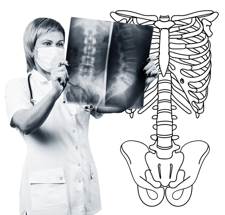
The diagnosis of lumbar spine osteochondry is based on clinical data and data for additional research methods.The key role belongs to methods such as:
- Radiography of the lumbar spine.
- Computational tomography of the lumbar spine.
- Magnetic resonance imaging of the lumbar spine.
Lumbar spine x-ray is necessarily performed in 2 mutual vertical projections-straight and side.Such images allow you to see the shape, outlines and structure of the vertebral bodies, the height and shape of the intervertebral discs, the spine abnormalities and the natural turns.To appear the intervertebral joints and the intervertebral holes, the radio lines are produced in oblique projections.To detect the pathological motility of the individual lumbar parts (which is an indication of osteochondrialism), radiography is performed under the conditions of functional test, that is, the bending and spinal extension.Normally, you can clearly see the change in the height of the intervertebral discs on the front or rear section according to the direction of the body slope, with osteochondria due to the operating block of one of the sections, the height of the disc does not change either during the bending or the farm.Pathological mobility determines the displacement of the vertebrae forward or back.The main signs of x -ray of osteochondalism include intervertebral slit narrowing, pathological mobility and shift of vertebral bodies, the deposition of salts in the disc (calcification), the formation of peripherals of the vertebral bodies,(calcification).Radiography of the lumbar spine is a research routine that gradually loses its importance in the context of active application of new and more informative research methods (CT and MRI).Lumbar x -ray is currently used as a diagnostic method of sorting.
The lumbar spine CT is also performed using X -ray radiation, but the radial load on the body is much smaller than with x -ray.The study is done on the table of a special device - a computer tomography, is absolutely painless.The resulting images are processed using a computer and allow you to see significantly more structures than with spine radiography.
Magnetic resonance imaging is a method in which electromagnetic radiation is used to create images.The study is also carried out in place to be on the table, which calls in the tomography chamber.Magnetic resonance imaging is harmless and painless.
CT or magnetic resonance imaging of the lumbar spine allow you to see all the spine structures, carefully examine the intervertebral discs (and the jacket and fibrous ring) and the intervertebral holes.Even a small protrusion of the intervertebral disc will not go unnoticed.These methods (especially magnetic resonance imaging) allow you to determine the direction of the disc hernia, if any, the degree of compression of the nerve roots, the spinal cord.Thus, these methods of research are much more informative in the diagnosis of the lumbar spine by radiography.In addition, they allow you to diagnose not only osteochondria, but also other diseases (tumors, spinal cord disorders, abscesses, congenital defects of spine structure and spinal cord), which is significant during the differential diagnosis.
The lumbar spine osteochondria is a disease that often causes back pain.It is, in fact, the destruction of intervertebral discs.Due to the osteochondicity of the lumbar spine of fate, a person often loses the ability to work, since, in addition to pain, the disease can lead to a violation of spine mobility, the inability to sit, stand and walk.Symptoms of this disease are non -specific and require additional research methods to accurately confirm the diagnosis.The most informative and safe for modern methods of diagnosis of osteochondry are the magnetic resonance imaging of the spine.